


 For Chemical Sciences With a Grant of Rs. 60 Lakhs")


Scientists from Paul Scherrer Institute, Switzerland, Friedrich-Alexander Universität Erlangen-Nürnberg, and other institutions in Basel, Hamburg, and Paris, have achieved placing a new record in X-Ray Microscopy. Using accurate sample positioning and advanced diffractive lenses, they were capable of succeeding single-digit nanometer-scale spatial resolution. This new finding in direct imaging could offer considerable momentum for studies of nanostructures and further ameliorate the creation of new kinds of magnetic data storage and solar cells. The study has been issued in the Optica journal with ‘Soft X-ray microscopy with 7nm resolution’ as the title.
Soft X-ray microscopy, which makes use of low-energy X-rays is utilized to check out the features of products in the nanoscale. This innovation can be used to identify the conformation of organic films that serve a crucial function in the growth of solar cells and also batteries. It also facilitates catalytic reactions
or chemical processes of products to be observed. The method enables the investigation of spin dynamics. New kinds of magnetic data storage can employ the electrons which can not only carry an electric charge but also possess an internal direction of rotation.To boost studies into these procedures in the coming years, scientists have to be capable of zooming in to the one-digit nanometre scale. This is possible in theory using soft X-rays, however, as of now, it has solely been able to succeed spatial resolution of less than 10nm with indirect imaging approaches that need further reconstruction. Prof. Dr. Rainer Fink, Chair of Physical Chemistry II, FAU elucidated that for active procedures like magnetic particle interaction or chemical reactions, they have to be capable of viewing the structures directly. X-ray microscopy is particularly apt for this because it can be employed more easily in magnetic settings than electron microscopy, for instance.
Improved calibration and focusing
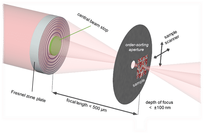
Combined works of researchers from institutions in Basel, Hamburg, and Paris, and Paul Scherrer Institute have hit the high spot in X-ray microscopy with their achievement of 7 nanometers, a record resolution, in various distinct experiments. This achievement is not rested chiefly on highly robust X-ray sources, but on ameliorating the X-ray focus with diffractive lenses and more accurate calibration of the test samples. Rainer Fink stated that they optimized the Fresnel zone plates’ structure size which is employed to focus X-rays. Further, they were capable of placing the samples inside the machine at relatively higher accuracy and generate this accuracy. It is obviously this restricted stability and positioning of the system as a whole that have avoided advancements in direct imaging resolution as of now.
Significantly, this fine resolution was not just accomplished with specially-designed examination structures, yet additionally in practical applications. For instance, the scientists examined the magnetic field alignment of iron materials determining 5 to 20 nanometres using their new optics. They hope that their outcomes will drive research further into nanomagnetism and energy materials in particular. The applicable structure sizes within these fields are commonly less than the present resolution limits, Prof. Fink clarified.
Author: Geema George


